FIGURE 3
.
Back to Liver Shunt article
.
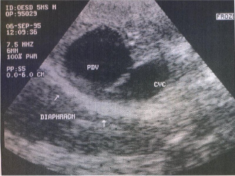
Ultrasound image of the liver from a dog with a portosystemic shunt. Note the
large anomalous patent ductus venous (PDV) adjacent to the caudal vena cava
(CVC) (photograph courtesy of P. Watson, University of Cambridge).
.
.